Managing the Gap With Immediate Implant Placement: Myths Versus Realities of Treatment

By: Dennis Tarnow, DDS
Managing the Gap With Immediate Implant Placement: Myths Versus Realities of Treatment
The placement of immediate implants has opened multiple discussions about how to manage the gap that may exist between the implant and the facial plate of bone. Some of the questions that should be addressed are as follows: Does the size of the gap make a difference in wound healing? Should a graft be placed into the gap? What graft material should be used? What is histologically present against the implant after healing? Does the graft change the outcome, or does it only act as a filler to maintain space and ridge shape? should the graft be covered with a custom abutment or a provisional?
1. Does the size of the gap make a difference in the wound healing?
This question emerged years ago as one of the key reasons many clinicians avoided placing implants into immediate extraction sockets. Animal studies showed that if the gap exceeded 1.5 mm, fibrous tissue could form against the facial aspect of the implant. However, closer review of those studies revealed a design flaw: after creating gaps of varying sizes, researchers achieved primary flap closure without placing a membrane (Ref. 1).
This allowed fibrous tissue from the inner flap to grow down along the implant before bone cells from adjacent walls could migrate and attach. Today, this concern has diminished because immediate implant placement into a fresh socket typically does not involve flap coverage. In fact, it is preferable not to elevate a flap at all.
This approach keeps epithelium and connective tissue away from the implant and allows time for bone to migrate to the exposed implant threads before soft tissue closes over the defect. Thus, contrary to earlier beliefs, the size of the gap does not matter (Ref. 2). It can be any distance, provided the socket heals by secondary intention rather than primary flap closure.
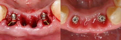
Notice that the space is larger than 2mm yet the healing went perfectly, even without a graft in the socket. Only a provisional was placed to keep the tissue shape. There was no probeable pocket depth where the gap was previously present.
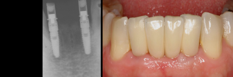
Final case in place along with radiographs (Restoration by Richard Smith)
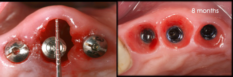
In this case, notice that there was a gap of 4.2 mm. Thanks to a gift from the patient, we were able to look at this histologically and show that the bone filled right up to the first thread as it would even if there was no gap present. This verifies the biological concept that gap size does NOT matter as long as there is no flap pulled over the gap. (Ref 2)

2. Should a graft be placed into the gap?
This is important because the graft seems to help preserve the contour of the ridge. The buccal bundle bone remodels, especially if it is thin. This is true almost 90% of the time after maxillary anterior teeth are removed. Thus, a graft should be placed, as most research shows that the contour of the ridge has less shrinkage with a graft than without a graft (Ref 3).
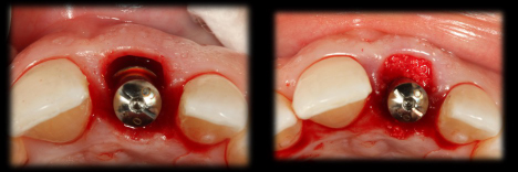
Graft placed into the gap
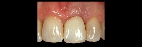
Provisional in place immediately after extraction and placement of the graft
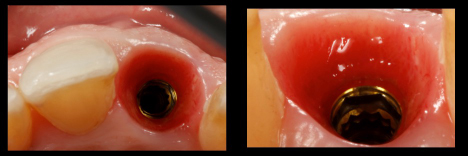
Wound healing following 4 months of healing with a provisional in place
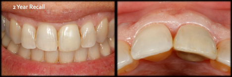
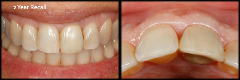
Final crown in place with 2-year follow-up. Notice that the tissue height and shape of the facial contour is barely changed with this process of grafting the gap.
3. What graft should be used if you do graft.
This is an interesting question that is mostly driven by companies instead of research. Many studies can be quoted to show that almost all grafts will help keep the shape of the ridge. Some may allow faster healing during the first two or three weeks, but there does not seem to be much of a difference after a period of a few months. If you want to make sure that the shape lasts, then use more nonabsorbable grafts, like bovine bone or calcified allograft. If you want more natural bone to be in the gap after healing, then use more absorbable graft material.
4. What is histologically up against the implant after healing? Does the graft change the outcome or does it only act as a filler to
maintain space and ridge shape?
This is something which many clinicians are not aware of. The reality is that whatever graft material you place, and no matter how firmly you place it, the particles are never against the implant after healing is complete. The graft is initially in a clot, and when angiogenesis occurs and new granulation tissue forms during healing, the graft particles are moved away from the bone. The particles are still in the gap, but they are NOT against the implant (Ref 4). This is true no matter what type of tissue or new bone ultimately forms against the implant. Thus, new bone, which is touching the implant, no matter how firmly the graft was placed. The graft material is still there if it resorbs slowly, but it’s within the new bone that formed in the gap between the implant and the old buccal plate of bone. (Ref 4,5)
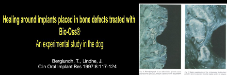
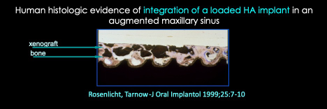
Notice that the bone graft particles in both studies show that the particles are NOT touching the implant after healing. The graft may still be present but not up against the healed implant surface.
5. Should the graft be covered with a custom abutment and/or a provisional?
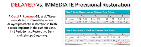
Evidence consistently shows that optimal esthetic outcomes are achieved when a provisional restoration, or at a minimum a custom healing abutment, is placed at the time of surgery. This supports soft tissue healing in the correct contour and minimizes collapse during the healing phase. Research demonstrates improved outcomes compared with sites left to heal without structural support for the gingiva (Refs. 3, 6). Additionally, the abutment or provisional helps contain the graft and stabilize it during early healing.
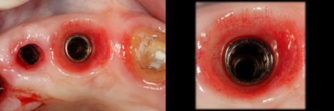
Notice that the tissue collapsed where there was no contoured healing abutment. However, notice the perfect shape of tissue right next to it when a custom healing abutment was placed. This saves the patient and the clinician chair time and at the same time give us better esthetic outcomes.
This 4-year longitudinal study shows that the vertical recession was about 1mm less if the provisional was placed immediately. The horizontal collapse was about 2mm more when the abutment wasn’t placed immediately.
Conclusions
This discussion illustrates an evidence-based approach leading to several key conclusions: The size of the gap does not significantly affect bone healing around an implant. Placement of a custom healing abutment or provisional helps maintain socket shape and minimizes contour shrinkage. Grafting the gap improves the ridge contour compared with leaving it unfilled. Graft material does not remain in contact with the implant after healing, regardless of type. The containment of the graft seems to help the shape and contour of the healed ridge.