Unforgiving Anatomy of the Socket Site: Are You Seeing What Matters?

Dr. Araujo
By: Maurice Araujo, DDS, MSc, PhD
Report from the Academy of Osseointegration 2026 Annual Meeting
During the Academy of Osseointegration 2026 Annual Meeting in Washington, DC, I presented an analysis of the biological and anatomical factors that govern tissue changes following tooth extraction and their implications for implant therapy. The presentation emphasized that
post-extraction remodeling should be regarded as a biologically driven process, the extent and pattern of which are largely dictated
by the anatomical characteristics of the alveolar socket.
The healing process at the extraction site follows a well established sequence, beginning with clot formation, progressing to the development of a provisional matrix, and culminating in new bone formation and maturation. However, these biological events inevitably result in a reduction of ridge dimensions due to the processes of bone modeling and remodeling. Consequently, even when an implant is placed immediately after tooth extraction in an ideal three-dimensional position, clinicians must recognize that the healed alveolar ridge will be diminished. This reduction can ultimately lead to esthetic challenges that must be addressed during treatment planning.
The presentation highlighted that these dimensional changes are not uniform across clinical scenarios. Instead, they are modulated by site-specific anatomical variables that have a marked effect on wound stability, the availability of bone cells, and blood supply. Among these variables, the thickness of the buccal bone wall appears to be particularly influential. Sites characterized by a thin buccal plate experience early buccal bone loss, which disturbs wound stability and leads to more pronounced horizontal contraction. In addition, the number and integrity of the socket walls, as well as the position of the tooth within the alveolar process relative to the basal bone, were identified as relevant determinants of healing outcomes.
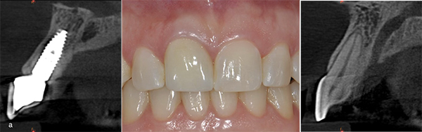
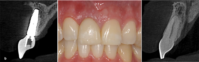
Figures: Left A, Right B
Two clinical cases exemplify the impact of gap and socket anatomy on alveolar ridge preservation following immediate implant placement. In case (a), a wide buccal gap (≥ 2 mm) was grafted at a site with a thick buccal wall. In case (b), the grafted gap was narrow (≤ 2 mm) and the buccal socket wall was thin. Both cases received a connective tissue graft (CTG) at the buccal aspect of the implants. Clinical and CBCT scans were obtained after 2 years of function. Notably, in the wide gap case, the ridge was successfully preserved, whereas a substantial reduction was observed in the narrow gap group.
These considerations have direct consequences for implant treatment planning. Although immediate implant placement has been proposed as a means of preserving ridge dimensions, available evidence indicates that it does not arrest the underlying remodeling process. As such, the decision to place implants immediately or to adopt a staged approach should be based on a careful assessment of the anatomical features of the extraction site rather than on timing alone.
Within this context, alveolar ridge preservation procedures were discussed as a strategy to mitigate, but not eliminate, post-extraction dimensional changes. By stabilizing the wound and compensating for buccal bone wall loss, the use of graft materials and barrier membranes may reduce the extent of ridge contraction. The effectiveness of these interventions, however, is closely linked to appropriate case selection and an accurate understanding of defect morphology.
The data presented, supported by both experimental observations and clinical examples, reinforce the concept that post-extraction tissue changes follow predictable biological patterns. Rather than attempting to prevent these processes entirely, clinicians should aim to anticipate them and incorporate this knowledge into treatment planning.
In conclusion, the lecture underscored the importance of a thorough anatomical evaluation of the extraction site as a foundation for decision-making in implant therapy. A biologically oriented approach to socket management remains essential for achieving stable and predictable outcomes, particularly in situations where esthetic demands are high.