
The eposteal implant: A new solution for the extremely resorbed ridge
By: AO Fellow and Diplomate Robert J. Miller, DDS
The endosseous root form implant represents a favorable option for tooth replacement in most cases of edentulism with very high success rates and predictable outcomes. However, endosseous implants require sufficient bone volume and quality to succeed, and these ideal parameters are not always available. In complex situations involving resorbed or compromised alveolar bone, the use of endosseous implants can be challenging or even impossible.
For almost a century, dental surgeons have attempted to devise strategies to stabilize dentures or other types of tooth replacements. Denture fabrication became an art form, with each iteration addressing a previous shortcoming in stability and comfort. Even with exquisite denture fabrication, further loss of the alveolus was unavoidable, and patients suffered the consequences of extreme alveolar bone loss (Figure 1).
The first attempt at fixation in the extremely resorbed ridge was the subperiosteal implant, designed to be placed over the existing bone volume with attachment points for a prosthesis. This implant was first introduced in Sweden by Gustav Dahl in the 1930s and then brought to the U.S. a decade later by Gershkoff and Goldman. It enjoyed a renaissance for another two decades as the preferred implant for the severely resorbed ridge. However, the absence of true hard tissue integration and a concomitant high failure rate resulted in this modality falling out of favor.
At the same time, the first endosteal (endosseous) implants fabricated in titanium drove the implant marketplace as a consequence of the concept of osseointegration. This type of implant remains the predominant class of implantable devices to date. While implant surgeons have become very adept at placing endosseous implants in a wide variety of cases, the extremely atrophic ridge remains problematic. Critical neurovascular structures, pneumatized sinuses, knife-edge ridges, and poor quality of bone remain limiting factors for these types of implants (Figure 2). Further, major bone grafting to create sufficient ridge volume is a time consuming process with considerable complexity, long healing periods, increased risk of infections, and limitations on vertical regeneration.
Consequently, surgeons developed additional implant modalities utilizing tilted, zygomatic, pterygoid, and nasalis implants to avoid grafting procedures and significantly reduce treatment time. However, these implants require a high level of training and carry much higher risks for potential bleeding episodes, neurosensory deficits, orbital perforation or infection.

Figure 1 – 3D modeling of a severely resorbed patient presenting for implant evaluation

Figure 2 – Panorex view for pre-treatment planning of eposteal implants
Approximately 10 years ago, clinicians in Europe started to revisit the concept of placing patient-specific implants over the existing bone complex to avoid bone grafting and potential injury to critical structures. In reviewing the literature, it was clear that the first problem they had to overcome was the lack of osseointegration as the result of using chrome cobalt materials. The choice was then made to use Titanium grade 5 alloy to ensure biocompatibility and even integration. With the proliferation of computerbased modeling, they no longer needed to take direct bone impressions in a first surgery and then deliver the implants at a subsequent appointment. The use of CBCT imaging with intraoral fiducial markers allows the surgeon to evaluate the planned prosthesis and then merge this plan with the current bone volume. The implants can then be virtually designed and printed with a choice of prosthetic attachments (Figure 3).
While all subperiosteal implants are eposteal, not all eposteal implants are subperiosteal as we may choose to graft over the implant. As a consequence, these implants may no longer be placed under the periosteum and therefore should not be classified as subperiosteal. We prefer to use the term eposteal implant as the major classification for any implant designed to be placed over the bone complex. It reflects our new concept of true integration of this type of implant modality.
Advantages of the eposteal implant include:
- Minimally invasive surgery that reduces risk to critical anatomy
- Fabricated in Sintered Laser Melted printed Grade V titanium alloy for strength and biocompatibility
III. Available in standard connections which include cementable, Morse taper, or multi-unit abutments
- Prosthetic connections that are placed in more ideal positions for ease of access to screw channels and stronger prosthesis
- Ideal force distribution as there are no cantilevered occlusal segments
- One-stage surgeries with immediate load options
VII. Biologic design for true integration to bone and preservation of soft tissue
VIII. Osseous graft options for covering the implant to enhance stability of the case long term
Over the past decade, eposteal implants have been used successfully to treat compromised patients with success rates rivaling that of endosseous implants. For the older patient who may be limited financially or have medical conditions that limit the extent of more complex surgical procedures that may be employed, the eposteal implant represents a realistic and literature-based alternative for the utilization of a stable and fixed implant-borne restoration.
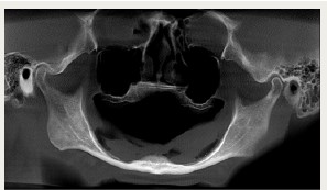
Figure 3 – Virtual implant planning for a segmented eposteal implant
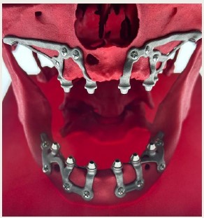
Figure 4 – Example of full-mouth eposteal implants for immediate load
References:
Mommaerts M. Evolutionary steps in the design and biofunctionalization of the additively manufactured sub-periosteal jaw implant ’AMSJI’ for the maxilla. Int J Oral Maxillofac Surg. 2019;48(1):108-114.
Cerea M, Dolcini GA. Custom-made direct metal laser sintering titanium subperiosteal implants: a retrospective clinical study on 70 patients. Biomed Res Int. 2018;2018:1-11.
Mangano C, Bianchi A, Mangano FG, et al. Custom-made 3D printed subperiosteal titanium implants for the prosthetic restoration of the atrophic posterior mandible of elderly patients: a case series. 3D Printing in Medicine. 2020;6(1):1-14.
Contact Information:
Robert J Miller, DDS
16244 S Military Trail, Suite 260
Delray Beach, FL 33484
Email: drrjmiller1@aol.com