
Operating microscope to improve predictability of periodontal and peri-implant therapy outcomes
By Albert Chan, DDS, MS
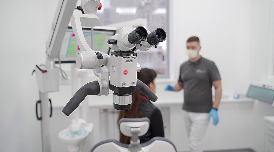
Technical advantages of the operating microscope (OM), including unparalleled magnification (up to 25x), coaxial illumination, and facilitation of the use of microinstruments as well as microsutures (6-0 and finer sutures), have been demonstrated in medicine and part of dentistry — for example, endodontics. The use of the OM is on the rise in the periodontal field. Taking Ohio State University (OSU), Ohio, as an example, our periodontal program is equipped with the OM for the residents to perform high-quality nonsurgical as well as surgical periodontal procedures.
Lectures as well as hands-on workshops pertinent to microsurgery are given on a regular basis to prepare our residents for having a minimally invasive mindset and precise periodontal care. Ultimately, the educational goal is to train our residents to become competent periodontal microsurgeons. As for the scientific rationale of applying the OM, accumulating evidence has shown its benefits at the tissue level and to some extent at the clinical level.
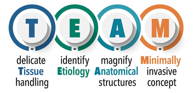
The acronym “TEAM” approach (Table 1) could further explain how the technical advantages can translate to clinical benefits with the use of the OM (DOI: 10.11607/ prd.2024.6.c). Successful implementation of the TEAM approach can ultimately contribute to improved clinical/patient-reported outcomes and operator well-being.
The explanation of the “TEAM” approach. The operating microscope allows for higher performance in these four key aspects.
Related to delicate Tissue handling, managing fine periodontal as well as peri-implant tissues is tactilely sensitive. The common goal is to exert as little trauma as possible while preserving tissue integrity, volume, and microvasculature (DOI: 10.1002/cap.10265; DOI: 10.11607/ prd.6213). This is especially true for tissue regenerative/reconstructive attempts.
It is also challenging to practice on thin phenotype, commonly characterized by narrow (less than 2 mm) keratinized mucosa, soft tissue of thickness less than 1 mm, and narrow interdental papillae (2-3 mm wide). High magnification provided by the OM facilitates seamless visual-tactile coordination. For example, during flap elevation, signs of incipient tissue tear under the OM can be captured, and the magnitude and direction of the force translated from the surgical elevator to the tissue then can be modified. This precise tissue handling practiced and monitored under the OM is a critical gateway to achieve predictable regenerative outcomes.
The use of OM makes it a breeze to identify and control/ eliminate the Etiology. A fundamental requirement to predictably treat implant-related conditions is to recognize and control or eliminate any etiological factors, such as microbial biofilm, calcified deposits, residual cements, and defective restorations. The current threat to implant dentistry is peri-implantitis, which compromises function and esthetics of a significant portion of the population with dental implants. Additionally, attempting reconstructive strategies to rescue implants is currently not predictable. The four interrelated negative conditions hampering effective tissue reconstruction are: inferior tissue perfusion, unfavorable bone topography, ineffective surface treatment, and unstable wound (DOI: 10.1002/ cre2.788). As the primary etiology is dysbiosis, implant surface decontamination is key to creating a biocompatible environment for tissue reconstruction. While biofilms might be relatively easy to remove, calcified deposits (necrotic bone or dental calculus) between the implant threads can be difficult. Under high magnification (~25x), surface decontamination can become more efficient, potentially resulting in improved outcomes.
The OM can magnify Anatomical structures — for example, mental nerve and the Schneiderian membrane — which are commonly encountered during implant-related surgeries. Surgical safety by avoiding damage to these sensitive structures is the priority. When releasing facial flap for hard/soft tissue regeneration in the mandibular premolar-first molar region, the mental nerve should be protected. On the other hand, being overly cautious could also result in inadequate flap releasing, negatively affecting the clinical outcome. Therefore, being able to visualize and navigate around mental foramen without damaging it could be achieved under the OM. In the same way, the integrity of the Schneiderian membrane should not be compromised in maxillary sinus augmentation (DOI: 10.11607/prd.5407). The OM is especially useful for investigating membrane integrity during vertical/ internal sinus augmentation (DOI: 10.1111/cid.13403). Early detection of membrane perforation can lead to proper damage-control strategies by, for example, containing the perforation size, not placing bone particulates, choosing a shorter implant, or aborting the surgery in extreme cases. Currently there is no adequate method to evaluate membrane perforation; therefore, the OM presents itself as an opportunity for this purpose.
The last word “M” relates to Minimally invasive approach. One of the main indications for surgery is to gain access. The traditional “one-size-fits-all” thinking for flap design and management may not be applicable anymore. Instead, a customized approach based on the surgical needs and bearing a concept of “start as small as possible, extend as large as necessary” in flap design should be implemented.
After all, any types of incision and flap reflection can cause disruption of tissue integrity, undesirable tissue trauma, compromise of microvasculature, and wound instability, which may result in the occurrence of mucosal recession, scar tissue formation, increased surgical time, and concomitant patient morbidity, among other possible adverse events. The advantage of the OM in this aspect is to allow for smaller access and at the same time achieve the same goal, compared with the traditional approach.
To summarize, the use of OM to improve surgical predictability is promising through the “TEAM” approach. In addition to the technology itself, deliberate practice and cognitive coordination between visual cues and tactile sensations are equally important. These technical along with nontechnical skills will eventually contribute to higher odds of surgical success that we are all longing for.